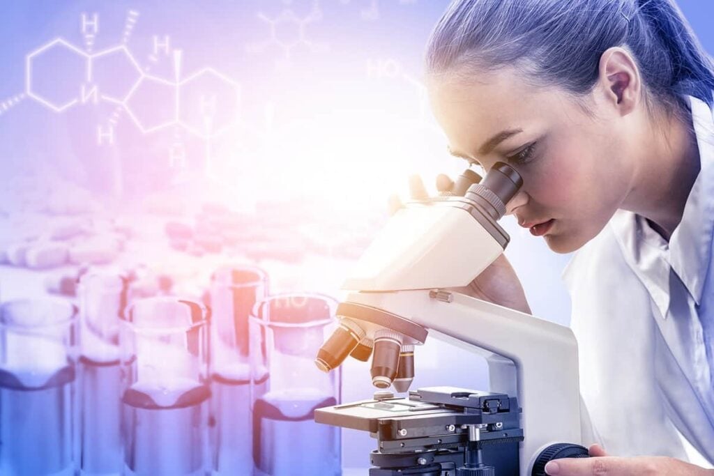
How Eikon Uses Fluorescence Microscopy for Drug Discovery


Table of contents

Back in the 1670s, a Dutch cloth merchant by the name of Antonie van Leeuwenhoek invented the first microscopic lens to get a better view of his cloth threads. Using his new invention, he discovered the microscopic world of cells, bacteria, and oddly enough, sperm. He singlehandedly became the father of microbiology. While it’s a bit tougher nowadays to land a second career as a world-class scientist after a stint in the fashion industry, advanced breakthroughs in microscopy are still making big splashes in the world of science. Much like how the first microscopes revealed a bountiful realm full of scientific possibilities, modern microscopes let us do so much more than just look at our fingernails up close or capture Instagram-worthy images.
What is Fluorescence Microscopy?
Fluorescence microscopy is one of the latest breakthroughs in the field of microscopy. Fluorescence is a fancy word that refers to substances that re-emit light after absorbing light of a different color. Just think of those scenes in forensics shows where the police stain and shine ultraviolet light onto a crime scene, and the bloodstains glow green. In fluorescence microscopy, cells or tissues are stained with a specific type of dye called a fluorophore, which re-emits colored light after being exposed to a certain wavelength of light in the form of a laser. The re-emitted light is a different color than that of the laser so that it can be filtered out for viewing. A camera is then used to capture the colored image that’s produced.

The fluorophore dye can be attached to specific molecules of interest, such as nucleic acids, proteins, or carbohydrates. The fluorophore can also be a fluorescent protein that’s genetically engineered to be produced by the cell itself. The technique allows scientists to create fancy images of the cell interior. Different structures and molecules can be stained with different fluorophores, creating a psychedelic pattern of shapes and colors that provide insight into the structure, mechanics, and movement of a cell. These scientific insights are still early days though, with the fluorescence microscopy market estimated to be worth $500 million back in 2018.

However, there’s a physical limit to conventional fluorescence microscopy. The resolution of the images is only as good as the wavelength of light that you use, and for light that you can see, that’s between 400 to 700 nanometers. It’s a lot like staring really closely at a flat-screen television and being able to see the individual pixels. Great for blown-up photos of colored cells, but what if a researcher wanted to look at one protein at a time? No such luck. Images are too blurry at that level of detail.
A Startup Peers Deep into Cells
Bay Area-based Eikon Therapeutics is a life sciences startup co-founded in 2019 by Eric Betzig, who won the Nobel Prize in Chemistry in 2014 for his work on super-resolution fluorescence microscopy, the 2.0 version of fluorescence microscopy. Eikon has raised $148 million in disclosed funding to date from the likes of Innovation Endeavors, the venture capital firm of Google’s (GOOG) former CEO, Eric Schmidt, after a Series A for that amount that was completed in early May 2021.
Update 01/07/2022: Eikon Therapeutics has raised $518 million in Series B funding to accelerate platform development and advance multiple discovery programs. This brings the company’s total funding to $666 million to date.
The company is building state-of-the-art tools to stare deep into the inner recesses of cells. Super-resolution fluorescence microscopy is to cell-based microscopy what quantum dot LED technology is for flat screens. That means smaller pixels, better contrast, and better color. Using complex math, multiple lasers, and multiple lenses, super-resolution fluorescence microscopy can capture sharp images of cellular objects as small as 100 nanometers. By taking microscopy to the next level, researchers can use Eikon’s visualization platform to track how small molecules, like proteins, shuffle around inside cells.
Proteins are some of the most important elements of the cell, doing pretty much all the work. That allows researchers to track the behavior and movements of these individual biomolecules inside the cell after specific biological events, such as those following treatment of the cell with a potential drug candidate.
Using Fluorescence Microscopy for Drug Discovery
Where proteins go and how they interact with one another are important insights for drug discovery. Imagine for a second that you’re taking aspirin for a throbbing headache. The aspirin passes through the stomach lining, where it travels into the bloodstream and finds its way to the brain. There, the drug gets absorbed by your neurons and jams up a protein in these cells called cyclooxygenase A, which is responsible for creating that headache in the first place. Along the way, there are proteins involved that facilitate the breakdown and transport of aspirin into the cells. There are also proteins that are produced by the cell to send signals to other parts of the cell and to neighboring cells after aspirin does its thing.

We only know this because of all the data collected from the thousands of experiments done to test different tissues, cells, enzymes, and other biomarkers. That data is good enough to make a very strong inference about what’s going on, but researchers never actually see these processes happening in a living cell. The situation is like the story of the blind men and the elephant, where each blind man feels a different part of the elephant thinking it’s a tree, snake, rope, or some other funky object depending on where they touched the elephant. But they never see the elephant itself. In the same way, scientists use limited information to piece together these different perspectives to create a theory on what’s going on inside the cell.
Eikon Therapeutics wants to do away with all that by using its super-resolution fluorescence microscopy platform to get a clearer picture of the cellular processes that happen when drugs are added to living cells. The platform relies on CRISPR gene editing to fluorescently label target proteins and track their location using super-resolution microscopy. This strategy produces terabytes of imaging data that researchers can use to understand the minute details involved in the cellular uptake, metabolism, and binding of a drug candidate. The data is then dumped into a machine-learning algorithm that tries to extract insights from that giant pool of information.

Super-resolution fluorescence microscopy allows Eikon to watch carefully where proteins go and what they do when a cell is dosed with a new potential drug molecule, providing higher-quality hard evidence on whether or not a drug candidate has a good chance at successfully passing the FDA’s intense drug-approval process. That means higher chances of success in an industry where experimental products experience greater than 90% failure and the mean cost of bringing a new drug through the development pipeline is about $1 billion. So far, Eikon has revealed a new hard-to-hit protein target involved in the progression of cancer that was previously believed to be undruggable, with three additional unnamed protein targets in the screening pipeline.
Conclusion
A picture might be worth a thousand words, but the right picture of a cell can be sold for thousands of dollars. And you know you’re in a great position as a startup when you have the Nobel Prize-winning inventor of the fluorescence microscopy technology you’re trying to sell on your team.
Eikon is creating a new window into the cell that’ll expand our ability to explore the previously unknown using some of the most cutting-edge technology available. Its platform will create new opportunities to capitalize on discoveries that’ll add buckets full of data to the drug development pipeline, and that means more cash in everyone’s pocket.
Share